Primary hyperparathyroidism is present in up to 0.1% of the general population, and it is clinically recognized in patients presenting with hypercalcemia or as a part of multiple endocrine neoplasia (MEN) types I and IIa. Although there have been sporadic reports of the coexistence of hyperparathyroidism & follicular adenoma/ thyroid malignancies, concurrence of parathyroid adenoma and follicular adenoma is rare.
In most reports discussing the coexistence of these two diseases, primary hyperparathyroidism was diagnosed before the identification of the thyroid adenoma/ thyroid malignancy, which is usually diagnosed in a pathology specimen.
Since the coexistence of both disease processes can complicate patient management. An untreated hypercalcemia & unrecognized thyroid adenoma/malignancy, there is necessity pre-operatively that the patients should be screened for both diseases carefully.
This case report highlights coexistence of parathyroid adenoma and thyroid adenoma (follicular) in a same patient & on same side of thyroid gland.
38 years old lady complaining of tiredness & lethargy since last 2 years along with history of renal stone disease. On investigation found to have hyperparathyroidism
PTH 240 pg /mL (15-65), Sr calcium 10.9 mg/dl (8.80 to 10.6 mg/dl). TFT- wnl.
Left lobe and isthmus appear to be enlarged. Evidence of hypodense nodule (26*20*12 mm) noted in the isthmus in the thyroid on left lobe, showing mild heterogenous enhancement in post contrast sections, lesion is causing focal anterior bulge with displacement of strep muscles.
A hypodense nodule noted in the lower pole of the left lobe of thyroid ( 15*10*8 mm), showing mild heterogenous enhancement in post contrast sections.
Evidence of oval shaped iso-dense area showing marked enhancement noted (30*10 mm) postero-superior to the left lobe of thyroid and shows marked enhancement in post contrast sections.
A focal mildly hyperdense nodule 10.8*7 mm noted in this area showing minimal enhancement in post contrast sections. with focal mildly hypodense nodule- possibility of parathyroid lesion. Right lobe appears to be normal.
Enlarged isthmus and left lobe of thyroid showing hypodense nodules. An oval shaped iso dense area showing marked enhancement noted (30*10 mm) postero-superior to the left lobe of thyroid and shows marked enhancement in post contrast sections, possibility of parathyroid lesion to be ruled out.
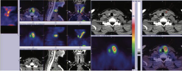
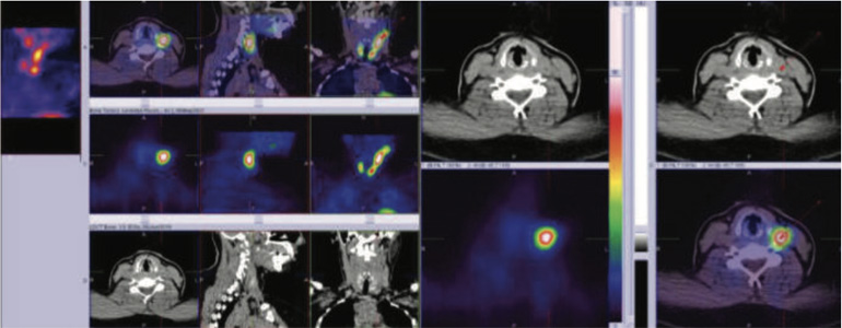
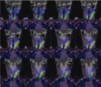
30 MINUTES HYBRID SPECT-CT images shows areas of increased tracer uptakes in a) lower third portion of left lobe b) upper half of the left lobe (larger one)- figure 2,3,& 4)
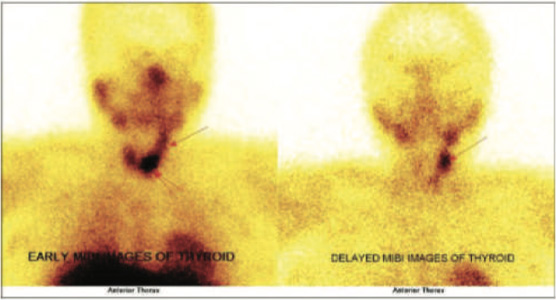
Delayed 2 hours images (washout images) shows persistent tracer retention in upper half of the left lobe hot area, while lower third left lobe hot area shows minimal tracer retention while rest of the thyroid shows adequate tracer washout pattern- figure
METABOLICALLY ACTIVE THYROID NODULE/ ADENOMA IN LOWER THIRD PORTION OF THE LEFT LOBE (EARLY HOT WITH MINIMAL TRACER RETENTION ON DELAYED WAHOUT IMAGES)-figure 1,2 & 4)
METABOLICALLY ACTIVE PARATHYROID ADENOMA IN UPPER HALF PORTION OF LEFT LOBE POSTERIORLY ( HOT ON SPECT-CT FUSED IMAGES AT 30 MINUTES AND PERSISTENT HOT ON DELAYED WASHOUT IMAGES)- figure 1,3 & 4)

Figure 1: 99mTc-Sestamibi early and delayed static images

Figure 2 - 99mTc-Sestamibi hybrid SPECT-CT (early) images showing focal area of increased tracer uptakes in lower third portion of left lobe lobe of thyroid gland (thyroid adenoma)

Figure 3 - 99mTc-Sestamibi hybrid SPECT-CT ( early) images showing focal area of increased tracer uptakes in upper half portion of left lobe lobe of thyroid gland (parathyroid adenoma)

Figure 4 - 99mTc-Sestamibi hybrid SPECT-CT( early) images showing focal area of increased tracer uptakes in upper half portion of left lobe lobe of thyroid gland (parathyroid adenoma)
Patient underwent left parathyroidectomy for primary hyperparathyroidism & left hemi thyroidectomy due suspicious thyroid nodule ( FNAC- suspicious for follicular lesion)
The final pathology revealed not only hypercellular parathyroid consistent with parathyroid adenoma (PTA) in the upper portion of the left lobe lesion and features of follicular adenoma in the lower 3rd portion lesion.
Since synchronous thyroid and parathyroid disease was first described in 1947, the incidence of thyroid disease in patients undergoing parathyroidectomy has been reported to be from 2.5 to 17.6%. Conversely, the frequency of primary hyperparathyroidism in patients with thyroid disease has been reported to range from 0.3 to 8.7%.
The frequency of coexistence of primary hyperparathyroidism and thyroid follicular adenoma is not well-known; In the present case, on 99mTc Sestamibi parathyroid scintigraphy, metabolically active parathyroid and thyroid adenoma has been suspected based on the scintigraphy pattern. The final diagnosis of concurrent parathyroid adenoma and thyroid follicular adenoma has been established on postoperative histopathologic examinations
In conclusion, although concurrent parathyroid adenoma and thyroid follicular adenoma is rare, they can and do coexist.