Increasing awareness of primary hyperparathyroidism screening may represent a key strategy for preventing recurrent kidney stones and other complications.
3% to 5% of patients with kidney stones have primary hyperparathyroidism (PHPT), which is a treatable cause of recurrent stones. However only 1 in 4 stone formers with hypercalcemia undergo testing for PHPT.
The apparent low prevalence of PTH testing in patient with kidney stones and hypercalcemia suggests low awareness among clinicians regarding this condition. We are missing an important opportunity to prevent recurrent kidney stones by diagnosing and treating PHPT. This is despite guidelines by the American Urological Association and European Association of Urology that recommend Calcium and PTH testing
Thirty-nine years old gentleman with past history of kidney stones four years ago which had passed after adequate hydration. At that time, he was found to have high calcium levels, but no further follow up was done. He did not report any renal episodes subsequently Recently he was admitted with hemoptysis and diagnosed as pneumonia (covid negative). Found to have a mass behind the thyroid gland
HRCT CHEST- Nearly complete consolidation of the left lobe. Mild pericardial effusion. Soft tissue mass measuring 26 x 15 x 25 mm located laterally to the upper thoracic trachea and posterior-inferiorly to the left lobe of thyroid gland? parathyroid nodule.
USG NECK- left infra thyroid paratracheal and esophageal loop related specific mass? atypical lymph node
FNAC of the nodule- Parathyroid tissue and possibly primary hyperparathyroidism (Adenoma).
Labs -Serum calcium 3.05 mmol/L (2.08- 2.65), PTH 18.50 pmol/L (1.95-8.49), TSH 1.14 (0.550-4.780 mIU/L), Vitamin D3 18.5 ng/ml, Sr creatinine 84 micromol/L (62-115), CRP 77.
Referred for 99mTc-SESTAMIBI parathyroid scintigraphy
99mTc-SESTAMIBI PARATHYROID IMAGES
EARLY AND DELAYED MIBI IMAGES OF NECK AND CHEST
It shows focal area of increased MIBI tracer uptakes ( approximately 2.0*1.6*2.2 cms in size) seen inferio-posterior from the lower pole of left lobe of thyroid gland on early images ( Better appreciated in SPECT-CT fused images) and persistent tracer retention is seen in the same of delayed washout images.
Thyroid gland shows comparatively decreased tracer uptakes on early images and adequate washout pattern on delayed images.
No focal area of any abnormal tracer uptakes seen in mediastinal region
The right lobe measures 4.4*2.0 cms in size and left lobe 5.2*2.4 cms in size approximately and good tracer uptakes.
THERE IS FOCAL METABOLICALLY ACTIVE PARATHYROID ADENOMA (approximately 2.0*1.6*2.2 cms in size) SEEN INFERIO-POSTERIOR FROM THE LOWER POLE OF THE LEFT LOBE OF THYROID GLAND ( figure 1,2 and 3)
Forty-five years old gentleman with recent history of renal colicky pain on left side. On investigations found to have ureteric stone for which he underwent lithotripsy. Besides this he also had a small stone in right kidney ( 2 mm).
Past history of left renal colic seven years back and found to have 14 mm stone, for which he underwent lithotripsy and became symptom free.
History of underlying diabetes mellitus, lipidemia, gout and on regular medications.
Investigations for recurrent renal calculi revealed high serum calcium and parathyroid hormone. Serum calcium 11.4 mg/dl (8.0-10.6), PTH 276.6 pg/ml (15.0-68.3), Serum creatinine 1.0 mg/dl (0.72- 1.18), Serum uric acid 6.4 mg/dl (3.5-7.2).
99mTc-MIBI PARATHYROID IMAGES
EARLY AND DELAYED MIBI IMAGES OF NECK AND CHEST
It shows focal area of increased MIBI tracer uptakes ( approximately 4.8 * 5.5 mm in size) seen mid medial portion of right lobe posteriorly on early images ( Better appreciated in SPECT-CT fused images) and persistent tracer retention is seen in the same region on delayed washout images (Better appreciated in SPECT-CT fused images)
Thyroid gland shows asymmetrical tracer uptakes (right lobe relatively > than the left lobe) on early images and adequate washout pattern on delayed images.
No focal area of any abnormal tracer uptakes seen in mediastinal region
The right lobe measures 4.5*2.5 cms in size and left lobe 3.8*2.0 cms in size approximately and shows asymmetrical tracer uptakes (right lobe relatively > than the left lobe).
THERE IS FOCAL METABOLICALLY ACTIVE PARATHYROID ADENOMA (APPROXIMATELY 4.8 * 5.5 MM IN SIZE) SEEN MID MEDIAL PORTION OF RIGHT LOBE POSTERIORLY (figure 1,2,3)
Unlike most recurrent stone formers, those with PHPT have a good chance at permanent cure. This makes detection of PHPT a paramount aim for patients and their physicians. PHPT results in elevated serum calcium and is detected by blood tests. The elevation of serum calcium can be slight and variable so patience and persistence matter a lot. Once diagnosed, PHPT can be cured by surgery that modern instruments and techniques have made very safe and highly successful in the hands of expert parathyroid surgeons.
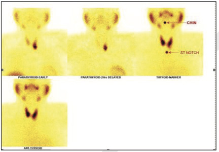
Figure 1
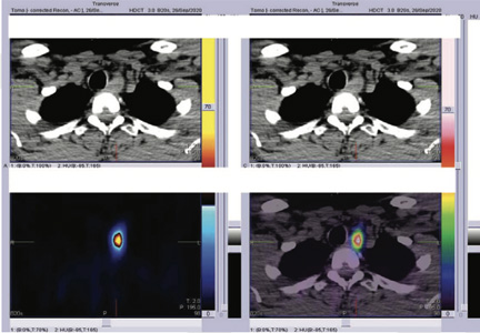
Figure 2
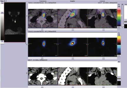
Figure 3
THERE IS FOCAL METABOLICALLY ACTIVE PARATHYROID ADENOMA (approximately 2.0*1.6*2.2 cms in size) SEEN INFERIO-POSTERIOR FROM THE LOWER POLE OF THE LEFT LOBE OF THYROID GLAND ( figure 1,2 and 3)
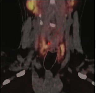
Figure 1 Early SPECT CT fused images
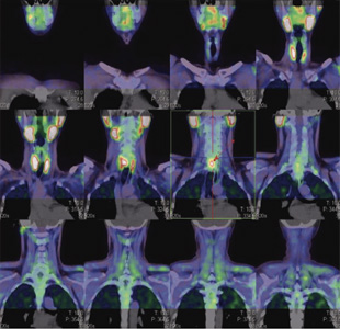
Figure 2- Early SPECT CT fused images
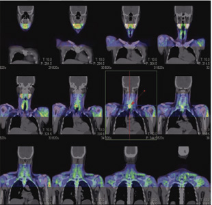
Figure 2- Early SPECT CT fused images