Neuroendocrine tumors include such tumors as adenomas from the pituitary gland, islet cell tumors from the pancreas, pheochromocytoma and neuroblastoma from the adrenal medulla, medullary thyroid carcinoma from the C-cells of the thyroid gland, carcinoid tumors from the gastrointestinal tract (or less often from the lung), and paragangliomas.
Generally, anatomic and/or physiologic-functional imaging for neuroendocrine tumors is reserved for patients who present with a clinical history that is suspicious for a neuroendocrine tumor, supported by elevated levels of primary hormones and/or metabolites in the plasma and/or urine. These patients can be evaluated by anatomical imaging studies, such as computed tomography (CT) or magnetic resonance imaging (MRI), and the functional status of these tumors can be assessed by physiologic imaging via scintigraphy, with radiolabeled octreotide (Octreoscan). These imaging techniques should be considered as complementary studies, rather than competitive modalities, as each provides important aspects in the evaluation of these underlying tumors for patient management.
In addition to localizing sites of disease, scintigraphy of neuroendocrine tumors can provide a functional assessment of the disease state. With whole-body SPECT-CT hybrid imaging capability, nuclear imaging is an excellent technique for searching for distant metastases or multifocal disease (as shown in this case)
Scintigraphy can also evaluate for tumor recurrence, for example, in evaluating abnormalities such as suspected scar tissue versus a recurrent tumor at or near a postoperative site and can also evaluate the efficacy of therapeutic interventions. Finally, scintigraphy can be performed to evaluate tracer uptake characteristics of a tumor when there is consideration of radionuclide therapy.
39 years old gentleman with history of recurrent epigastric pain, anorexia, weight loss and jaundice.
Large pancreatic mass lesion in head/uncinate process of pancreas of around 75*58 mm with grossly dilated Common bile duct, common hepatic duct with dilated IHBR with few ill-defined focal lesions 17*12 mm in segment 8, suspicious of metastasis.
Large pancreatic mass lesion in head/uncinate process of pancreas of around 63*53*46 mm with grossly dilated common bile duct, common hepatic duct with dilated IHBR with mildly dilated pancreatic duct. Few ill-defined focal lesion, largest 15 mm seen in segment 8 suspicious of liver metastasis. Common bile duct is compressed by the mass in the distal end close to the ampulla.
Underwent endoscopic ultrasound guided biopsy of the mass.
large ulcerated necrotic heterogenous subepithelial duodenal mass involving second/third part of duodenum.
Histopathology/immunochemistry revealed Grade II Neuroendocrine Tumour
Referred for whole body octreoscan (99mTc Tektrotyed) for the staging of the disease process
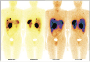
99mTc-TEKTROTYED WHOLE BODY IMAGES
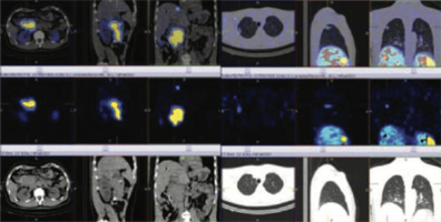
SRS LESION IN LARGE DUODENAL MASS SRS LESION IN RIGHT UPPER LUNG ( METS).
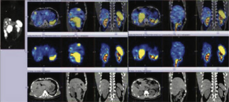
MULTIPLE SRS LESIONS IN LIVER ( METS).
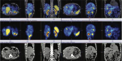
MULTIPLE SRS LESIONS IN LIVER ( METS).
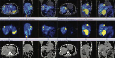
MULTIPLE SRS LESIONS IN LIVER ( METS).
99mtc- labelled-tektrotyed {hynic octereoscan whole body scan (somatostatin receptor imaging)}.
H/o large duodenal mass involving second/third part of the duodenum & biopsy suggestive of grade 2 neuro-endocrine tumor.
Somatostatin receptor expression (srs) lesions are seen in:
All abnormalities are better appreciated on spect-ct fused images
What is the clinical relevance of somatostatin receptor scintigraphy?
A positive scan predicts the suppressive effect of somatostatin analogues on hormone secretion from endocrine active tumours.
The 99mtc-tektrotyed whole body and spect/ct hybrid imaging has high sensitivity, and it makes it a suitable, easily accessible nuclear imaging method to detect neuroendocrine tumors and staging of the disease process as highlighted in this case.