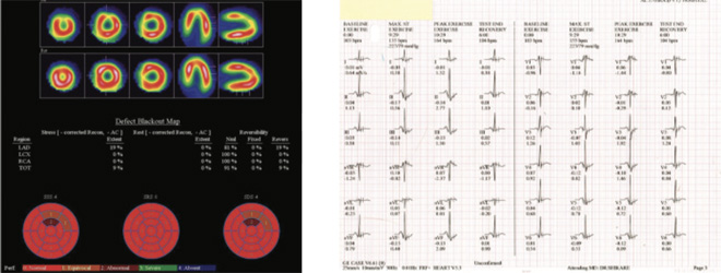
34 yrs old male presented with left sided chest pain radiating to the back- one episode. No h/o diabetes, hyperlipidemia, smoking , obesity, hypertention and any family history of IHD. Pulse- 91 Bpm, bp 119/84 mm of Hg, 2D Echo- WNL
Treadmill exercise with Bruce protocol- exercise time 10.29 minutes, work load achived 10.60 mets, Maxmimum heart rate 164 bpm and BP 227/79 mm of Hg, 87% of maximum age predicted heart rate. Exercise stopeed-THR achived with no symptoms.
EKG showed no significant changes.
MILD REVERSIBLE PERFUSION ABNORMALITY IN APICO-ANTERIOR AND PORTION OF ANTERIOR WALL OF THE MYOCARDIUM. MINIMAL REVERSIBLE PERFUSION ABNORMALITY IN LATERAL WALL ( DISTAL 2/3RD) AND SEPTAL WALL ( DISTAL 1.3RD), APICO-INFERIOR AND INFERIOR WALL CLOSE TO APEX OF THE MYOCARDIUM AT 87.0% OF PMHR (PREDICTED MAXIMUM HEART RATE) AND 10.60 METS. QUANTITATIVELY
The apico anterior and portion of anterior wall perfusion abnormality corresponds to LAD TERRITORY and is of minimal in nature ( 19%) with 19% reversibility. Rest of the perfusion abnormality as mentioned above are not quantitatively significant.The approximate total myocardium under ischemic risk is 9% with 9% reversibility.
All arteries were normal and right coronary were dominant
As per the literature- Ischemia on MPI (stress myocardial perfusion imaging) is predictive of long term adverse cardiovascular outcomes despite of normal coronary angiography ( Normal is defined here as no visible disease or luminal irregularities (< than 50%) as judged visually at coronary angiography).
There is strong correlation between the regions of the original perfusion abnormality and the ultimate coronary ischemia or revascularization.
Abnormal findings on MPI may predict a higher prevelance of coronary and peripheral vascular events than suggested by a normal coronary angiogram.
Perfusion imaging unlike angiography, closely correlated with flow reserve and reflects function of epicardial conduit arteries as well as normal capacity of the resitance vessels.


