To observe the renal function recovery measured by diuretic renography in short term follow up of patient with transperitoneal laparoscopic pyeloplasty (Anderson–Hynes)
The ureteropelvic junction obstruction (UPJO) in adults is defined as a functional disorder of the trans-port of urine from the renal pelvis into the ureter, producing pyelic and calyceal dilatation of the affected kidney. Although the most common cause is a congenital alteration of the ureteropelvic junction, in which the obstruction is due to intrinsic fibrosis of a ureteral segment that becomes aperistaltic, other causes have been described from a ureteral origin (ureteral valves, anomalies in the insertion of the ureter) to extraure-teral (adhesions, fibrosis, abnormal vessels)..Multiple surgical techniques have been described, having evolved considerably over the past 20 years with the advance of new technologies. Traditionally, treatment of UPJO was based on surgical open pyeloplasty.
Anderson and Hynes open pyeloplasty consists mainly of the ablation of the ureteral stenosed segment, the removal of part of the dilated pelvis and the performance of a ureteral muco–mucosal anastomosis. The effectiveness of open pyeloplasty approaches 90%, but carries with it significant post-operative pain and a prolonged hospital stay.In recent years, many minimally invasive techniques have been developed; the laparoscopic and endoscopic approaches of UPJO have reduced the morbidity of open surgery. Laparoscopic pyeloplasty obeys the same rules as the open surgery described by Anderson–Hynes, benefiting from the advantages of minimally invasive surgery. The intervention may be performed by transperitoneal or retroperitoneal approach with similar results to open surgery ac-cording to several published series.
Now a days the assessment of overall renal function, the ureteropelvic anatomy and physiology is considered essential in the diagnostic process of UPJO, since these parameters determine the appropriate choice of the surgical technique to use. Diuretic re-nography is the most commonly used diagnostic tool to assess for UPJO.
Technetium Tc 99m MAG3 (Mercapto Acytyle Glycine) is the radiopharmaceutical agent of choice for this purpose and has largely replaced Tc 99m Diethylene Triamine Penta- acetic acid (DTPA), because it has a better gamma image than DTPA, as well as a faster clearance rate and lower background activity. One advantage of DTPA is that it may be used to measure the glomerular filtration rate, but MAG3 scanning may provide differ- ential renal function by comparing isotope uptake in both kidneys, which, in turn, is a reflection of renal blood flow.
The aim of this study is to observe the renal func-tion using diuretic renography in short and medium follow–ups of patients after laparoscopic pyeloplasty.
Thirty one year old gentleman with history of left sided pelvi-ureteric junction obstruction causing left sided moderate pelvi-calictasis and slight parenchymal thininning on contrast CT scan of kidney, ureter and urinary bladder.
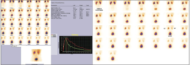
Ref for 99mTc MAG3 Diuretic renal scan for further work up 99mTC MAG3 DIURETIC RENAL SCAN (I/V LASIX AT 20 MINUTES) (pre operatively)
Left Kidney: Moderate but maintianed cortical functions (37.8%). Dilated pelvi-calyceal system with partial moderately severe obstructive uropathy pattern at UPJ post diuresis (peaking time 10.5 min (3-5 min) and T ½ of excretion not reached (10-12 minutes) ) and % of tracer retention at 30 minutes is 73% approximately.
Left kidney renogram curve shows moderate parenchymal phase with prolongation of peaking time with slow and delayed excretory phase, which has further improved after diuresis
Right Kidney: Good cortical functions (62.2%) with non obstructive tracer excretion pattern.
Right kidney renogram curve shows good parenchymal with non obstructive excretory phase.

Fig 1 & 2 - 99mTcMAG3 Diuretic Renal scan and Renogram (Pre-operative)
Subseqently Underweight Left Laproscopic Pyeloplasty in September 2020
Refered for follow up 99mTc MAG3 Diuretic Renal Scan for Reasseesmnet of Differential Renal Functions and to confirm obstruction has been resolved post operatively (Twelve weeks)
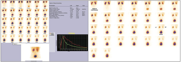
99mTC MAG3 DIURETIC RENAL SCAN (I/V LASIX AT 20 MINUTES). (Post operativey)
Left Kidney: Good cortical functions (50.5%). Prominent / minimally dilated renal pelvis with non-obstructive tracer excretion pattern (peaking time 4.o min (3-5 min), T 1/2 of excretion 12.5 min (10-12 min) , % tracer retention at 30 minutes 13% approximately.
Left kidney renogram curve shows good parenchymal with relatively slow and delayed but non obstructive tracer excretion pattern as compared to opposite kidney.
Right Kidney: Good cortical functions (49.5%) with non obstructive tracer excretion pattern and the % tracer retention at 30 minutes 13% approximately. Right kidney renogram curve shows good parenchymal with non-obstructive tracer excretion pattern.

Fig 3 & 4 - 99mTcMAG3 Diuretic Renal scan and Renogram (Post-operative)
Laparoscopic pyeloplasty not only corrects the UPJO, it also may recover renal function demonstrated after twelve weeks of follow up with diuretic renography. Laparoscopic pyeloplasty should be procedure of choice even in those patients with poor renal function at diagnosis, whenever there are chances of recovering renal function, regardless patients age.