Osteitis pubis (OP) is a debilitating overuse syndrome andcharacterized by pelvic pain and local tenderness over the pubic symphysis commonly encountered in athletes often involved in kicking, twisting, and cutting activities in sports such as soccer, rugby, football and to a lesser degree distance running. It is a common source of groin pain in elite athletes attributable to pubis symphysis instability as the result of microtrauma caused by repetitive muscle strains on pubic bones. Diagnosis is based mainly on detailed sports history and a meticulous clinical examination, although occasionally is difficult to distinguish this entity from other pathologies affecting the involved area which may occur concomitantly in the same patient.
Muscle imbalances between the abdominal and hip adductor muscles have been suggested as an etiologic factor in osteitis pubis. Because of their attachments to the thoracic cage proximally and the pubis distally, the abdominal muscles act synergistically with the posterior paravertebral muscles to stabilize the symphysis, allowing single-leg stance while maintaining balance and contributing to the power and precision of the kicking leg. The adductors, because they stabilize the symphysis by bringing the lower extremity closer to the pelvis, are antagonists to the abdominal muscles. In addition, the adductor muscle group transmits mechanical traction forces toward the symphysis pubis during its activity as a prime mover in the soccer push pass, tackling, and directing the soccer ball. Imbalances between abdominal and adductor muscle groups disrupt the equilibrium of forces around the symphysis pubis, predisposing the athlete to a subacute periostitis caused by chronic microtrauma. This microtrauma exceeds the dynamic capacity of tissue for hypertrophic remodeling, resulting in tissue degeneration. Shear stress at the symphysis pubis can also cause sacroiliac dysfunction in osteitis pubis if hip internal rotation is limited in either flexion or extension. This shear stress is transmitted to the symphysis pubis, resulting in either anteroposterior movement of one half of the pelvis in relationship to the other in extension or proximal-distal movement in flexion. The incidence of this syndrome in the general athletic population has been reported as 0.5% to 6.4%.
The clinical diagnosis of osteitis pubis was based on the history, physical examination, and radiographic (x-ray examination and bone scan) findings. Because the origin of groin pain is often difficult to elucidate, the sport medicine team must consider several differential diagnoses; thus, the players were examined by the orthopedic surgeon and a general surgeon to eliminate inguinal hernia, urologic pathologic conditions, muscular injury, or other orthopedic problems.
23 years old gentleman with history of severe pain in the lower abdomen for three weeks. It started as acute onset after playing football, when he slipped and fell (professional football player). Since than unable to play the football anymore and had difficulty in walking. On examination severe tenderness in symphysis pubis.
X-rays of the pelvis- shows picture of irregularity in the symphysis pubis with lytic lesion on left side. MRI of both hip joint- shows grade I strain of the left obturator externus and right gluteus maximus muscle
Referred for the 99mTc-MDP bone scan and Spect CT of Pelvic Region with Suspicion of Pubic Periosteitis.

Figure 1: CT Scan images of Pelvis shows (figure 1)
Joint cavity is maintained, articular margins are irregular, and sclerosis is note in subchondral bone on either side (left side is more prominent and associated with osteolytic changes involving the superior pubis ramus.

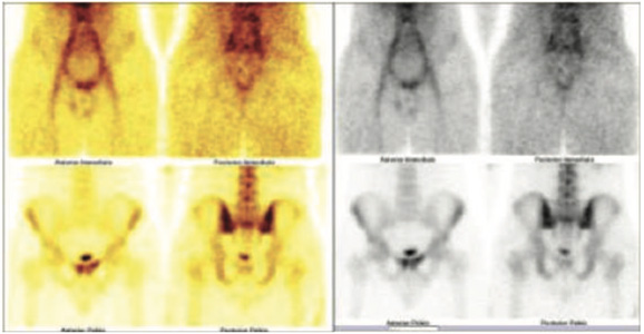
Figure 2 :99mTc-MDP Blood pool and bone images of pelvis ( figure 2)
It shows abnormal pooling of the tracer and abnormally increased tracer uptakes in the symphysis pubic region extending to the superior pubic ramu on left side medially.

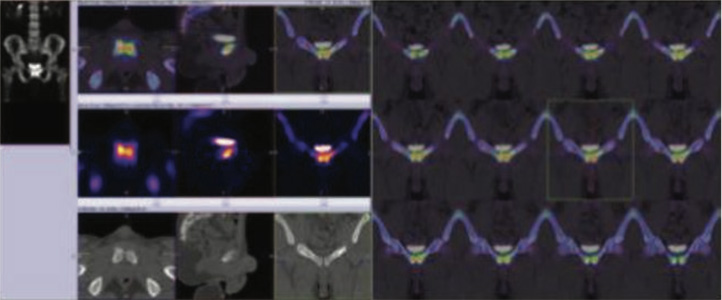
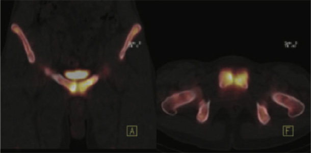
Figure 3

Figure 4
Spect CT Fused Image of Pelvis (figure 3 & 4)
It shows abnormally increased tracer uptakes in the symphysis pubic region extending to the superior pubic ramu on left side medially.
A high degree of suspicion for the presence of osteitis pubis (OP) should be raised at the clinician involved in the investigation and management of groin pain in the elite athletes with repetitive strenuous activities on pubic bones and the surrounding soft tissues. Early recognition of this ailment is imperative to avoid mismanage and to secure early and uneventful return to full sports activities. OP is usually a self-limited pathologic condition and most of the cases recover spontaneously or respond well to conservative treatment.