Metabolic bone disease encompasses several disorders that tend to present a generalized involvement of the whole skeleton. The disorders are mostly related to increased bone turnover and increased uptake of radiolabeled diphosphonate. Skeletal uptake of 99mTc-labelled diphosphonate depends primarily upon osteoblastic activity, and to a lesser extent, skeletal vascularity. A bone scan image therefore presents a functional display of total skeletal metabolism and has valuable role to play in the assessment of patients with metabolic bone disorders.
Currently, the main clinical value of bone scan in metabolic bone disease is the detection of focal conditions or focal complications of such generalized disease, its most common use being the detection of fractures in osteoporosis, pseudo fractures in osteomalacia and evaluation of Paget’s disease. 99mTc diphosphonate bone scanning agents Phosphate analogues can be labelled with technetium (99mTc) and are used for bone imaging because of their good localization in the skeleton and rapid clearance from soft tissues, and their uptake in bone is thought to reflect osteoblastic activity and to a lesser extent skeletal vascularity.
Osteoporosis is characterized by a decrease in bone mass with thinning of the cortex and trabeculae and a reduction in the number of trabeculae. Bone densitometry is the most diagnostic procedure for the detection of reduced bone mass. The bone scan has not been found to have an important role to play in the diagnosis of osteoporosis.Loss of vertebral height and closeness of the rib cage to pelvis in patients with multiple vertebral fractures may be observed in bone scan. These features are not diagnostic, but their presence may alert on the presence of osteoporosis. In practice the bone scan provides a less reliable means of diagnosing osteoporosis than radiography. Osteoporotic bones are abnormally brittle and are at high risk of fractures from mild trauma. These are easily recognizable on the bone scan and are focal areas of increased tracer uptake. If vertebral collapse is present, the classical appearance of a fracture in bone scan is horizontal linearly increased tracer uptakes.
Patient with history of backache & osteoporosis with vertebral collapse seen on x-rays. Whole body Bone scan show linear shaped increased tracer uptakes, typical of benign vertebral collapse (fig 1- L4 and L5 vertebrae & fig 2- L1 vertebra).
THE CLASSICAL APPEARANCE OF A FRACTURE ON BONE SCAN IS HORIZONTAL LINEARLY INCREASED TRACER UPTAKES ( FIG 1 & FIG 2) & CAN BE USEFUL FOR VERTEBROPLAS- TY FOR TREATING SPINAL FRACTURES DUE TO OSTEOPOROSIS (HOWEVER COEXISTING PATHOLOGY IF ANY, CAN NOT BE EXLUDED ON THE BASIS OF BONE SCAN ALONE ).
Osteomalacia results from vitamin D deficiency, which produces a profound mineraliza- tion defect. A common etiology is a lack of vitamin D caused by different factors. In severe cases there is massive excess of osteoid present with markedly reduced mineralization. The bone scan appearances in osteomalacia are usually abnormal. The scan findings, however, are non-specific and will demonstrate the classical characteristics of metabolic bone disease. Generalized increased uptake, most visible at the periarticular zones, costochondral junctions, vertebral column, calvaria, mandible, and sternum. Bone uptake of tracer reflects skeletal metabolism and that is likely to be primarily due to parathyroid hormone effect Pseudo fractures (Looser’s zone or milkman’s fractures) are often detected early in the disease, and are usually symmetric and perpendicular to the bone surface. They are commonly found in the scapula, medial aspect of the femoral neck, pubic rami, ulna (proximal third), radius (distal third), ribs, clavicle, metacarpals, metatarsals, and phalanges. Pulsating vessels on the softened bone cortex have been suggested to be the origin of pseudo fractures. Pseudo fractures are most often seen in the ribs. It is uncommon to see pseudo fractures in isolation at other sites such as femoral neck or pelvis. The bone scan provides a sensitive means of identifying pseudo fractures, particularly in the ribs, where conventional radiology cannot detect the lesions.
Whole body bone scan shows High tracer uptakes in axial skeletal, high tracer uptakes in long bones, high uptakes in peri-articular areas, faint or absent kidney images, Beading of costo-chondral junctions, tie sternum. Multiple scattered hot spot in ribs (pseudo fractures), suggestive of Metabolic bone scan disease (fig3)
Paget’s disease of bone is a common disorder in the elderly in which excessive production of structurally abnormal bone occurs. Paget’s disease is usually polyostotic but may be monostotic. It is characterized in its initial phase by excess resorption of bone, which is followed by an intense osteoblastic response, with deposition of collagen in a mosaic pattern rather than the lamella arrangement seen in normal bone. Paget’s bone is extremely vascular, and it has been suggested that this contributes to the bone pain. It is usually asymptomatic and normally is discovered incidentally after the detection of an elevated serum alkaline phosphatase, or a finding on X-ray or bone scan. The most common sites are the pelvis, spine, femur, tibia, and the skull, but any bone in the skeleton may be affected. The weight-bearing bones were most often symptomatic, lesions which appear normal radiographically but abnormal on the bone scan are generally asymptomatic.
The bone scan appearances in Paget’s disease are often characteristic, with predominant feature being markedly increased uptake of tracer throughout most or all of the affected bone. Lesions in Paget’s disease may be clearly identified because of the clear delineation between normal and abnormal bones. Bone scan is recognized to be more sensitive than radiography in detecting sites of Paget’s disease in the skeleton, with the additional advantage of visualization of the whole skeleton in one exploration. Paget’s disease usually affects transverse spinous processes of the vertebrae. The affected vertebra has then a typical appearance that has been described as a “Mickey Mouse’’ sign because of the inverted triangular uptake distribution. Other typical signs that have been described are the “black beard” sign (monostotic mandible disease) and the “short pants” sign (spine, pelvis and upper femoral disease) . The bone scan is generally of value in detecting complications of Paget’s disease such as fracture or sarcoma. In Paget’s sarcoma, there is usually no evidence of increased uptake at the site of developing malignancy, unlike in uncomplicated Pagetic bone. Distinction between Paget’s disease and metastases is usually easy on bone scan when a typical Paget’s disease pattern is present. A single lesion on the bone scan is generally more difficult to interpret, but even in this situation the typical scan features are likely to differentiate monostotic Bone scan may be a better diagnostic indicator of long-term response than biochemical measurements. Bone scan is the most sensitive technique for Paget’s disease detection and staging and should be used in primary evaluation. Because of its high sensitivity and the possibility to check the whole skeleton in the same study, bone scan should be the imaging technique of choice, with radiography used to obtain supplementary information whenever required. In clinical practice, bone scintigraphy is the ideal screening test in cases of suspected Paget’s disease and in elderly patients with unexplained bone pain, bone deformity, or in patients with an elevated serum alkaline phosphatase.
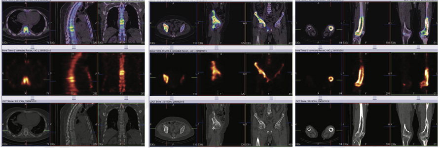
History of leg pain for one year, along with prostatitis. X-rays, CT and MRI of left leg suspected to be giant cell tumor with increased vascularity. Whole body bone scan shows abnormally increased tracer uptakes in right hemi pelvis and proximal third of left tibia favoring Polyostotic Paget’s disease (fig 4-left row).
Biopsy: Paget’s disease. Follow up whole body scan on treatment shows significant improvement ( fig 4- right row)
53 years old male, suspected case of paget’s disease of skull with neurological deficit. Bone scan: Monostotic paget’s skull bones.
Fig 5A- Whole body bone images show abnormally increased uptakes in skulls bones. Rest of the skeletal is unremarkable on whole body images favoring monostotic paget’s disease.
Primary hyperparathyroidism is a common disorder involving an increased secretion of parathyroid hormone that causes hypophosphataemia and hypercalcaemia. Most cases are mild and asymptomatic, but in advanced cases, peptic ulcer, pseudogout, nephrolithiasis and less frequently, excessive bone resorption can be present. Radiographic changes are not usually present because the disease is diagnosed with increasing frequency and at an earlier stage. Bone scan usually shows slight generalized increased uptake with high bone to soft tissue ratios. The degree of bone scan abnormality generally reflects the amount of skeletal involvement.The bone scan is the more sensitive of the two investigations and if scan appearances are not suggestive of metabolic bone disease then radiographs will invariably be normal [26]. A comparison between visually graded 99m Tc-HEDP images for metabolic characteristics and radiography showed that the first one detected 7 of 14 studied patients with primary hyperparathyroidism, whereas the latter detect 3 of them [26]. Despite apparently normal diphosphonate uptake on the bone scan by visual assessment, quantitative measurements (e.g. 24-hr WBR of diphosphonate) are frequently elevated as a result of mild diffuse increase in skeletal metabolism [1]. Ectopic calcification of the lungs and stomach can occur in advanced hyperparathyroidism, and can be detected with radiophosphate imaging [30]. Ectopic calcification is not a diagnostic feature; it can be present after the use of cytotoxic drugs, malignancy and in other conditions producing hypercalcaemia. The presences of focal abnormalities on the bone scan in primary hyperparathyroidism are uncommon but may be seen when Brown tumours are present, with chondrocalcinosis, or following vertebral collapse. In rare instances when a patient presents with aggressive, rapidly advancing primary hyperparathyroidism, multiple sites of ectopic calcification may be seen on the bone scan.
Bone scintigraphy plays an important role in the management of metabolic bone disease. It’s most important features are the high sensitivity, the capacity to image the whole body and the capacity to show typical diagnostic patterns of abnormality. Metabolic bone diseases in bone scan is highly sensitive method for demonstrating disease in bone, often providing earlier diagnosis or demonstrating more lesions than are found by conventional radiological imaging.
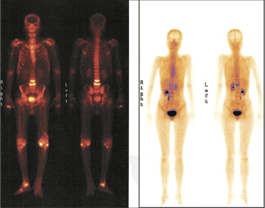
Figure 1- L4 and L5 vertebrae, Figure 2 - L1 vertebra
The classical appearance of a fracture on bone scan is horizontal linearly increased tracer uptakes ( fig 1 & fig 2) & can be useful for vertebroplasty for treating spinal fractures due to osteoporosis (however coexisting pathology if any, can not be exluded on the basis of bone scan alone ).
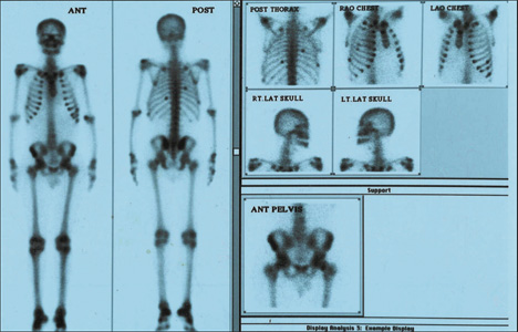
Fig 3: Metabolic bone scan features as seen in above case High tracer uptakes in axial skeletal, high tracer uptakes in long bones, high uptakes in peri-articular areas, faint or absent kidney images, Beading of costo-chondral junctions, tie sternum. Multiple scattered hot spot in ribs (pseudo fractures).
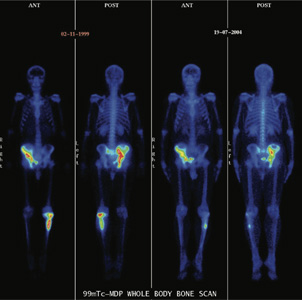
Fig 4 99mTc MDP WHOLE BODY BONE SCAN Whole body bone images shows abnormally increased tracer uptakes in right hemi pelvis and proximal third of left tibia favoring Polyostotic Paget’s disease ( left row). Biopsy: Paget’s disease
Follow up Whole body bone scan (right row) on treatment shows significant decreased in tracer uptake pattern in left tibial abnormality and mild to moderately decreased in tracer uptake intensity in right hemi pelvis lesion
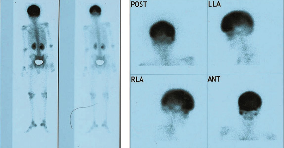
53 yr old male,Suspected case of paget’s skull with neurological deficit. Bone scan: Monostotic paget’s skull bones.
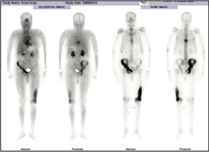
65 yrs male with h/o severe dorsal spine pain since 2 months. MRI spine- Suspicion of D8-D9 infective Spondylo discitis with canal stenosis. BONE SCAN – Abnormal uptakes involving D8 & D9 vertebra with destruction of disc space ? Infective. Abnormal right pelvis and left femur uptakes favors polystotic pagets Biopsy- Dorsal vertebral region-Tubercular.
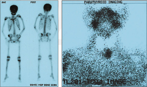
17 yr old girl with h/o fall, sustained #lt. humerous thought to be pathological. Bone scan shows features of metabolic bone disease. Sr. Ca High, Low Phosphorous, PTH: elevated Tc-Tl parathyroid substraction scan shows presence left lobe parathyroid Adenoma. Sx. Confirm the same.