Injury of the pars interarticularis is one of the most common identifiable causes of ongoing low back pain in adolescent athletes. It constitutes a spectrum of disease ranging from bone stress to spondylolysis and spondylolisthesis. Bone stress may be the earliest sign of disease. Repetitive bone stress causes bone remodeling and may result in spondylolysis, a non-displaced fracture of the pars interarticularis. A fracture of the pars interarticularis may ultimately become unstable leading to spondylolisthesis.
The differential diagnosis for back pain is broad and includes degenerative disease, infection, inflammation, tumors and trauma. Injury of the pars interarticularis is one of the most common identifiable causes of ongoing low back pain in adolescent athletes. It constitutes a spectrum of disease from bone stress through spondylolysis and spondylolisthesis. Bone stress may be the earliest sign. It is most common at L5, which is particularly vulnerable to micro-trauma from repetitive flexion, extension or rotational forces. Repetitive bone stress may result in spondylolysis, a non-displaced fracture of the pars interarticularis. Ultimately spondylolisthesis, or slippage of one vertebral body on another, may occur.
Radiographs of the spine have limited sensitivity compared with other imaging modalities in detecting bone stress and acute spondylolysis. Furthermore, radiographic defects of the pars interarticularis may not be symptomatic
Single photon emission computed tomography (SPECT)/computed tomography (CT) is ideally suited for assessment of low back pain in children and young adults. Spondylolysis is one of the most common structural causes of low back pain and is readily identified and characterized in terms of its chronicity and likelihood to heal. The value of SPECT/CT extends to identification and characterization of other causes of low back pain, including abnormalities of the posterior elements, developing vertebral endplate, transverse processes, and sacrum and sacroiliac joint. Some of the disease processes that are identifiable at SPECT/CT are like those that occur in adults (eg, facet hypertrophy) but may be accelerated in young patients by high-level athletic activities. Other processes (eg, limbus vertebrae) are more unique to children, related to injury of the developing spine.
12 years old boy with 8-month history of low back pain. He is regular football player. Suspected spondylosis.
Ref for 99mTc-MDP BONE SCAN

It shows focal area of increased tracer uptakes in left L5 vertebra in the region of pars intercularis (break in pars intercularis of L5 vertebra)- Spondylolysis of L5 vertebra left side.

15 years old boy with12-month history of low back pain.
He is regular athlete and football player.
Suspected spondylosis.
Ref for 99mTc-MDP BONE SCAN
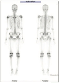
99mTc MDP whole body bone images It shows small focal area of abnormally increased tracer uptakes in L5

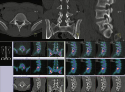
BONE SPECT CT FUSED images of lumbar region Focally active abnormality seen in pars intercularis of L5 vertebra on left side favors spondylolysis.
Low back pain occurs in 40% of children and adolescents at some point before adulthood. The prevalence of low back pain increases with age, becoming similar to that of adults by adolescence. Despite the frequency of low back pain, a structural cause is identified in only 12%–26% of cases.
When a structural cause for low back pain is identified, spondylolysis is commonly implicated since the pars interarticularis is the weakest portion of the immature neural arch.
Technetium-99m (99mTc) methylene diphosphonate (MDP) bone scintigraphy is an important tool in imaging workup of pediatric low back pain, particularly because of its utility in assessment of spondylolysis SPECT/CT has several advantages including more precise localization of sites of abnormal uptake in bone, identification of causes of abnormal uptake, and identification of osseous abnormalities without associated abnormal radiotracer uptake.